Swollen Joint, N.wheeleri....what is causing this, HELP?

Geckoholic

New member
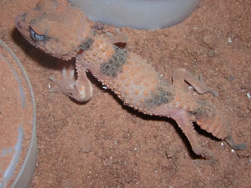
Let me start off by saying I had this exact problem with a taenicauda about 3 months ago. The gecko showed signs of a swollen joint on the front leg. She was brought to the vet and it was thought that she was suffering from septicemia, a bacterial infection (very possibly pseudomonas), and was given a total of 7 daily injections of baytril. The vet believed that a very small cut on the toe of that same leg (only really noticeable with the aid of a magnifying glass) was what started the infection. After 7 days there had been absolutely no decrease in swelling so the animal was brought back to be re-examined. I was then given a different antibiotic to be injected every other day (don’t remember what this was off the top of my head). After 2 additional weeks no progress was seen and there was now swelling at the joint in both font legs and one rear. Needless to say the animal ended up dying.

I am now seeing the exact say problem in one of my wheeleri. I do not want to see a repeat of the above incident. What is causing this swelling and how do I fix it. Any help will be GREATLY appreciated.

Thanks,
Steve


swollenjointwheeleri.jpg
 

Ari

New member
Hi

Just curious is it actually a joint swelling or is it fluid build up, i.e. fluid retention? Are the stools typical?

Cheers
 

Geckoholic

New member
Stools appear normal, in fact everything seems absolutely normal besides the obvious swelling. It is probably fluid build up but it is only seen at the joint.

Steve
 

Marlies

New member
I have an baby R. ciliatus that seems to have the same problem. The animal is feeding, can still climb the wall and seems to do OK, besides the swollen leg. I have no clue at all what the problem might be, but I'm going to follow this topic with great intrest!
 

oli

New member
I wish you all the best with your beautiful wheeleri. Someone with knowledge please help........
 

peterc

New member
Hi - an interesting problem you have there...some additional history may be useful, so how old is the wheeleri, was it sudden onset, is there lameness associated with the swelling, is it painful on palpation ( be gentle ),is it firm, was a PM performed on the last lizard...if so findings,any difficulties shedding...??????? Without detailed history and clinical examination +/- approp. tests I can only guess at the problem but from past experience the most likely cause of the problem would be one of the following osteomyelitis (bone infection),septic arthritis (joint infection), abscess, fracture with subsequent callus formation, or gout (common frillies and beardies..I haven't had a case in geckos). My approach would be a detailed radiograph using mammo. film and after reviewing that would consider performing a fine needle aspirate of afffected area and see what that yielded diagnostically. I assume wheeleri OS are expensive so could justify spending some money on vet workup to accurately Dx problem. Hope this is of some help Dr Peter Cameron. Hi to Dr Danny Brown...hope to meet you some time and chew the fat with you.
 

Geckoholic

New member
Thanks for the help Dr. Cameron. I will try to answer your questions as best I can...

The wheeleri is approximately 6 months old and the swelling appeared quickly. In fact, it now appears that one of the rear joints is starting to swell slightly (which is the same pattern seen in the taenicauda). The leg seems to be fully functional at this point but the animal doesn't seem to want to put a ton of weight it. The swollen area feels pretty hard, but it is hard to tell with such a small animal. There have been no shedding problems and the gecko is still feeding.

If it helps, here is how I care for gecko: 1 inch of sand/clay substrate, shoebox size enclosure with a partially screened top. 3 plastic saucers turned upside down, which serve as hide boxes. A corner of the enclosure is very lightly misted once a night and a heavier misting is done on half the enclosure once a week. I do not keep a moist hide with them as I do my levis. Half of the enclosure sits on a heat cable warming up the warm spot to the high 80's while keeping the cool section in the 70's. The wheeleri is fed exclusively crickets, which have been gutload (which does not contain animal protein) and dusted with calcium and vitamins (1 part repcal with D3, 1 part repcal without D3, and 1 part herpivite).

Let me know if there are any other questions that you would like answered.

Thanks,
Steve
 

Ari

New member
Good points there Peter. Actually have you found that shedding problems is the first visual sign of more severe underlining problems?

I had an adult female Pilbarensis ( 1 yr old ) she went into preshed, but stayed like this for 2-3 weeks, which concerned me, finally after taking her to the vet it seems she had past shed build up on one of her tiny toes which caused the toe to drop off and thus caused an infection - the leg showed signs of swelling and a antibiotic was administered orally but did no good. Poor thing passed away. This indicates to me that maybe when reptiles have major problems shedding that there is an underlining problem - I guess similar to cats in away, "wont clean its self if its ill?"
 

peterc

New member
Geckoholic...please call me Peter - if another joint is involved I would be more suspicious of septic arthritis, with spread via blood to other joints/organs.......normally however we would see other systemic signs/changes such as ventral erythema,petechial haemorrhages present oral mucosa, weight loss, anorexia etc. So it is interesting that the wheeleri is eating well etc. Reptiles are generally tough, hardy creatures ( there are obvious exceptions ie laevissimus) so you may still have a very sick gecko that is doing what it is programmed to do....fighting for survival and trying to get to reproductive size so that it can pass on its genetic material.Did you have a PM performed on the other gecko, if not did you open the affectede areas for your own interest, if so what did you find. If it is a septic arthritis are there any wounds on the gecko that may explain how bacteria entered, is the gecko housed alone?(Bite wound??)I suppose gout is still poss. DDx secondary to renal disease...has this gecko been treated with any drugs...if so what were they? If yes was the other gecko treated with same drugs ?A detailed radiograph would be useful (need right equipment and exposure techniqu though)...was the treating vet experienced with reptiles?

Ari...Hi there....not necessarily so........I always like to rule out poor husbandry (heating,lighting,diet,supplementation,humidity,substrate etc etc) first as usually the fault lies there!! Retained shed on extremities and subsequent loss of nail/toe is extremely common in shinglebacks (if I had a dollar.......) and bluetongues (often called constricted toe syndrome) bacteria then enter and infection sets in. bone destruction follows (lysis) and then lizard has a date with Dr Amputation......haven't seen this in geckos but I see relatively few geckos compared to other reptile species.
 

Geckoholic

New member
Thanks for the help Peter.

Although the wheeleri is currently eating I am skeptical that it will continue to do so. When I first noticed these symptoms in the taenicauda it too was eating. That same day I brought the gecko to the vet and from that point on the animals healthy steadily declined. I believe the gecko only ate a few times after that. Because the animal had been in good health up to that point it was able to survive for over 6 weeks. After 3-4 week 1 joint was swollen to an extreme and the other 2 were also swollen very badly. It lost the use of both back legs by the 3rd week and the animal’s spine also began to look odd. I thought for sure it would die within days. I had just ran out of injections at this point and didn’t see a point in acquiring more because the animal hasn’t been responding them and the damage up to this point looked irreversible. In addition all of that the tissue on the rear legs began dying by the 5th or 6th week. Would an extreme case of septic arthritis cause all of these things to happen? Unfortunately, the gecko died while I was out of town for a few days so I was unable to have a PM performed. How do you prevent septic arthritis when from my understanding it is initially contracted through commonly found bacteria?

The taenicaudea had a small wound on one of its toes that appeared to have possible stemmed from a “ring” of skin that had not come off during its last shed (other then a very small piece of skin on the animals back there were no others signs of a bad shed). I am unable to find any wounds on the wheeleri and it is being housed alone so another animal could not have inflicted an injury. The wheeleri (nor the taenicauda have ever been treated with any type of drug.) From what I hear the vet I take my geckos to is supposedly pretty knowledgeable, however, none of the above mention diagnostic techniques were applied.

Thanks again Peter.

Steve
 

peterc

New member
Geckoholic ( good name...an affliction shared by many on this site!!! )....it is looking more like a septic arthritis (hopefully Danny will have other insights) so how are bacteria entering? I reread all the posts to see if I had missed anything and some thoughts follow......the substrate looks coarse/abrasive...is it?Are there lots of sharp edges to the sand mix?Perhaps your humidity levels are too high...perhaps too frequent spraying.....I have never kept these species however. Is there anything else abrasive in cage?Some comments I made about generalised septicaemia (ventral erytheme,petechial haemorrhages)apply more commonly to snakes, turtles and less commonly to lizards...they were general comments and on rereading I felt I needed to correct statement.

Ari....any change from the norm may be an early indicator of a developing problem.....so record keeping is impt. - sorry gtg...more later. Peter
 

peterc

New member
Ari - as I was saying, any change from normal behav. MAY be important...things like loss appetite, shedding probs/freq, abnormal behaviour, calm animal becoming timid etc etc etc.........so record keeping is impt. Once your collection size increases then often accurate record keeping goes out the window as time is precious to all of us...which is a pity as there are huge knowledge gaps waiting to be filled. Peter
 

Ari

New member
Geckoholic it seems that the S. Taenicauda had some type of infection - how far gone it went is unknown. I wouldn't automatically believe that the Wheeleri has the same complications though - maybe, maybe not.

However if an antibiotic is administered make sure the correct dosage is given based on the size & weight of the specimen - as an over dose maybe more detrimental.

As per Peters previous recommendations related to the substrate - I agree and would convert to a much finer grade, and not as coarse and abrasive.

My Pilbarensis female's infection was noted by me far to late, it is correct to notice any changes in the specimens behaviour earlier if the case maybe - time restrictions.

I must say that was my first casualty and since then I have had no further problems. "Touching wood"
 

peterc

New member
If the wheeleri dies make sure you have a PM performed...if there are no gross lesions to explain problem then have histopath done - and incude whole affected limb/joint.Store in fridge not freezer prior to getting it to vet.If it is obviously going to die consider euth and PM to ensure best chance of correct diagnosis.Good luck and keep us informed - Peter.
 

Geckoholic

New member
Luc- I can’t be certain that the antibiotics didn’t play a role in the animals demise. I am sure the daily injections didn’t help either….

Peter: The substrate is a mixture of Jurassic red sand (which can be purchased at most local pet shops) and red clay which I collected locally from an area being developed here is SC. The mixture was an attempt to give the knob tails a harder surface which would be more natural and would also prevent easy ingestion of the substrate. With the amount of digging knob tails do, the sand alone was always breaking up even after the surface was misted and allowed to dry. However, keep in mind, when the taenicauda got sick it was before I made this change in substrate. At that point I was using straight Jurassic sand.

I only lightly mist one corner of the wheeleri cage each night (by light I literally mean 1-2 seconds). Approximately once a week 1 half of the tank is more heavy misted so that the substrate on that 1 side is damp for 1-3 days under the hides (not on the surface).

Ari- I agree, this could be something completely different then the taenicauda. As far as administering the antibiotics, all of the syringes are made prior to my leaving the vet so they should all be correct…at least I would hope…What substrate are you using Ari?


Vet Results…

I took the wheeleri to the vet yesterday. I was told by the vet that based on the animal itself and its age he doubted it was gout. I mentioned much of the advice I received here on the forums (thanks guys) and advice I have received viva e-mail. Such advice being a a radiograph and having the joint aspirated. He decided the animal was too small to have the joint aspirated. He thought the best course of action would be to lacerate the joint that was severely swollen and “drain it” by pushing on the swollen area. In the process he swabbed a portion of the “fluid” and is going to have it sent off and cultured in order to try and find out exactly what we are dealing with. He also wanted to get the animal stated on antibiotic right away. The wheeleri was given a shot of clafrin (which was the other anitbiotic used on the taenicauda) last night and I am to give him/her another injection tonight and then once every other night for a total of 7 injections…Lastly, some of the material extracted from the joint was gram stained. The results of such staining show numerous cells (which he said reinforced it was not gout) and only a few Gram positive cocci. No GNRs were seen.
 

Ari

New member
Hi Geckoholic

I tend to generally use double white washed sand mixed with coir peat on some species and then a red clay sand mined locally on others. I think you maybe ok with what you are using. I always have 1/4 of the enclosure's substrate moistened - whether they use it everyday or not is irrelevant in my eyes at least its offered for them to choose.

The humidity in my enclosures fluctuates between 60 - 75%. I reacon one very important thing to remember inrelation to a specimens recovery is less stress is best, personally administering any type of medication by injection cant be to good on such a small specimen. I prefer an oral substitute.

I wish you all the best with this little fella you have. Its imperative that we all take note of the gecko's toes when it comes to shedding as this can be easily missed but in both my case and yours it has had detrimental effects. Such a small small thing like a cut on a toe can easily be missed.
 

peterc

New member
Geckoholic - my strongest suspicion has always been an infectious cause.....I only included gout on mt differential diagnosis (DDx) list for completeness......note that it was listed last on list. My main reason for even including it was a concern re. overdosage of nephrotoxic drug (potentially kidney harming).
 
Top